

>>Nano-Cyte®
Additional Information
Brochure
Nano-Cyte® Video
Related Products
RM21™ Microscope Platform
C-Focus™
Nano-LPS Series
MicroStage Series
Nanopositioning Accessories
Questions?
E-Mail Us
 |  3D stability for imaging |
| US Patent Number 9,019,363 |
What is Nano-Cyte®? | |||||||||||||||||||||||||||||||||||||||||||||||||
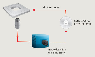
The Nano-Cyte® works by using the image of fluorescent fiduciary references, sparsely distributed within the sample, to localize these emitters in all three dimensions. The Nano-Cyte® uses this 3D localization information to provide active position adjustments to the sample, thus eliminating drift in the experiment. Nano-Cyte® is the complete stabilization and image acquisition instrument for advanced fluorescence microscopy. Our integrated approach to 3D stabilization yields image stability up to 3nm in X,Y and Z axes. Nano-Cyte® has proven stability over days and is a unique offering that promises to revolutionize advanced microscopy methods. | |||||||||||||||||||||||||||||||||||||||||||||||||
Hardware | |||||||||||||||||||||||||||||||||||||||||||||||||
The Nano-Cyte® is comprised of a high performance three axis piezo nanopositioning system coupled with a two axis motorized micropositioning stage. These precision motion capabilities enable the active positional control and particle tracking features of the Nano-Cyte®. The nanopositioner is a flexure guided piezoactuated design with integrated PicoQ® sensors for absolute position sensing and nanometer precision under closed loop control. The micropositioning stage enables the user to have a large range of travel for surveying samples prior to engaging the active stabilization. All motion devices are controlled by the Nano-Cyte® controller via USB 2.0 interface. The Nano-Cyte® hardware is compatible with Mad City Labs RM21™ open microscopy platform and most models of inverted optical microscopes. | |||||||||||||||||||||||||||||||||||||||||||||||||
Method | |||||||||||||||||||||||||||||||||||||||||||||||||
| |||||||||||||||||||||||||||||||||||||||||||||||||
Software | |||||||||||||||||||||||||||||||||||||||||||||||||
The native Nano-Cyte® software performs 6 important functions:
The Nano-Cyte® 3D stabilization occurs simultaneously with image acquistiion and incorporates reference selection, reference localization, and calibration statistics. Acquired images are saved in TIFF format and can be exported to ImageJ and other 3rd party software for post-acquisition processing. Nano-Cyte® device control ensures precision motion control and the ability to incorporate a variety of external user devices such as EMCCD cameras, shutters and light sources. Compatibility with LabVIEW™ and μManager facilitates even greater user device control and flexibility. Post-acquisition features of Nano-Cyte® enable the localization of particles within an image and the three dimensional rendering of particle positions. Nano-Cyte® is compatible with LabVIEW™, μManager, ImageJVI and rapidSTORM. In addition, Nano-Cyte® has an exportable DLL to allow wider functionality with 3rd party software platforms. | |||||||||||||||||||||||||||||||||||||||||||||||||
Stability Data | |||||||||||||||||||||||||||||||||||||||||||||||||
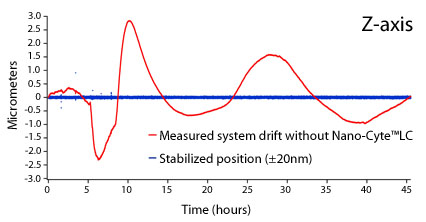
Nanometer Stability over DaysStability data measured over 44 hours for the Z-axis. The red line indicates the measured drift. The blue line indicates the stabilized position when using Nano-Cyte®. Similar results were observed simultaneously for the X and Y-axis. These data below demonstrate the efficacy of the Nano-Cyte® under non-monotonic drift conditions.
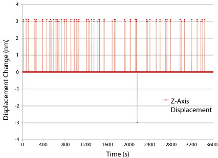
Effective Stability Using Nano-Cyte®The Nano-Cyte® data below shows position changes performed on the z-axis over a period 1 hour. Similar results were observed simultaneously for the X and Y-axis. These data demonstrate that the Nano-Cyte® effective stability is 3nm.
| |||||||||||||||||||||||||||||||||||||||||||||||||
Nano-Cyte® Specifications | |||||||||||||||||||||||||||||||||||||||||||||||||
| |||||||||||||||||||||||||||||||||||||||||||||||||
Additional Information | ||
Nano-Cyte® Brochure |
| |
|
Technical notes about sample preparation and applications are available by request. |
||
References | ||

 Powered by Bioz
Related Products |
Copyright © 2024